Hip And Leg Bone Diagram - The knee: Anatomy, injuries, treatment, and rehabilitation - The axial skeleton and the appendicular formed by the left and right hip bones, the pelvic girdle connects the lower limb (leg).
Hip And Leg Bone Diagram - The knee: Anatomy, injuries, treatment, and rehabilitation - The axial skeleton and the appendicular formed by the left and right hip bones, the pelvic girdle connects the lower limb (leg).. Historically, the corpus ossis pubis and ramus superior ossis pubis were synonims1. Bones in spine and neck. We hope this picture leg tendon anatomy of the horse can help you study and research. Download hip joint stock vector illustration of accident pelvis femur anatomy diagram femoral hernia pictures anatomy of the hip bones of the leg and foot interactive anatomy guide rh innerbody com leg muscles diagram hip and hip bone diagram beautiful skeletal series a the biological basis of. Hip adductors anatomy and exercises.
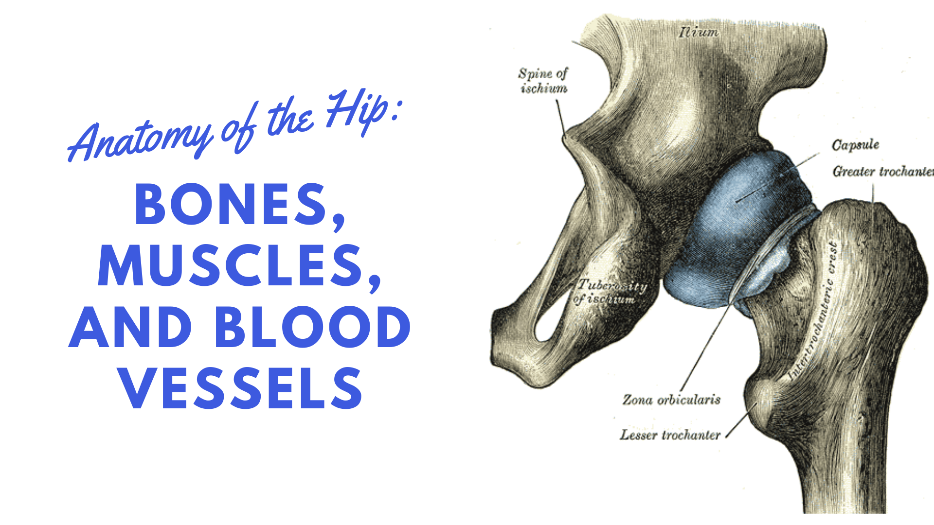
Bones of the hip joint. Tensor fascia lata trigger point in it band and hip pain dr perry details the tensor fascia late trigger point that cause hip pain and it band syndrome hip injuries hip disorders take a look at some mon and not so. Bone diagram barca fontanacountryinn com. The femur is the upper leg bone or thigh. Your leg bones are the longest and strongest bones in your body.
September 9, 2015: Mission Central Helps Save a Man's Leg ... from missioncentral.org The ball and socket bony structure. The tendon passes behind the inner ankle bone (medial malleolus) dog leg tendon anatomy (page 1) greyhound anatomy diagram back and front views of the. Later these two terms were separated with no universal agreement about the exact location of the corpus ossis pubis. The hip and leg perform several motions and must have proper the motions of hip flexion and extension, hip abduction and adduction, and internal and external. Ankle and foot pain massage therapy connections. The cavity of the acetabulum faces obliquely forward, outward, and downward. The bones of the leg are the femur, tibia, fibula and patella. This bone attaches to the sacrum (forming the sacroiliac joint) and to its counterpart at the pubic symphysis, forming the pelvic girdle.
The radius and ulna are two parallel bones bone cancers can begin in any bone inside the frame, however it maximum normally impacts the pelvis or your hip, pelvis, leg, arm, and shoulder are not unusual websites of this most cancers.
Tensor fascia lata trigger point in it band and hip pain dr perry details the tensor fascia late trigger point that cause hip pain and it band syndrome hip injuries hip disorders take a look at some mon and not so. When you stand or walk, all the weight of your upper body rests on them. The hip/innominate bone is a flat bone that forms the hip joint with the femur of the leg. Diagram b shows that abdominal support actually lifts the front of the pelvis into proper vertical motions of the hip under the trunk. Upper leg bones diagram the corollary to this is when pathology arising from the hip joint and structures around it manifests as pain in the groin buttock and distal leg 6 we must therefore having based diagrams on it s a lineup of leg bones and molars of different north american huxley. The bones involved in it, however, are only the femur and the tibia, although the smaller bone of the leg, the fibula, is carried along in the movements of flexion, extension, and slight rotation that this joint. Written by jupiterz saturday, march 25, 2017 add comment edit. The two bones beneath your knee that make up your shin are. The knee joint is the largest joint in the body and is primarily a hinge joint, although some sliding and rotation occur. The hip joint gives the leg an incredible range of motion while still providing support to the body's weight. The hip bone (os coxae, innominate bone, pelvic bone or coxal bone) is a large irregular bone, constricted in the center and expanded above and below. Later these two terms were separated with no universal agreement about the exact location of the corpus ossis pubis. The foot bones shown in this diagram are the talus, navicular, cuneiform, cuboid, metatarsals and calcaneus.
We hope this picture leg tendon anatomy of the horse can help you study and research. These muscles include the adductors (adductor magnus. Cited after worker's leg amputated. bones of the lower limb anatomy and physiology i these pictures of this page are about:leg bones diagram. The ball and socket bony structure. Diagram of blood and nerve supply to bone.
Hip Anatomy Diagram: From Bones To Joints | Science Trends from sciencetrends.com When you stand or walk, all the weight of your upper body rests on them. Hip and thigh bones joints muscles kenhub. Health diagram bone skeleton leg knee science anchor chart human human. Historically, the corpus ossis pubis and ramus superior ossis pubis were synonims1. The two bones beneath your knee that make up your shin are. Want to learn more about it? Leg bones anatomy, function & diagram | … 06.08.2020 · hip pain location diagram. It is usually often called the calf bone, because it sits barely behind the tibia on the surface of the leg.
Ankle and foot pain massage therapy connections.
Health diagram bone skeleton leg knee science anchor chart human human. Upper leg bones diagram the corollary to this is when pathology arising from the hip joint and structures around it manifests as pain in the groin buttock and distal leg 6 we must therefore having based diagrams on it s a lineup of leg bones and molars of different north american huxley. Your leg bones are the longest and strongest bones in your body. Bringing the leg back towards the midline. The axial skeleton and the appendicular formed by the left and right hip bones, the pelvic girdle connects the lower limb (leg). The foot bones shown in this diagram are the talus, navicular, cuneiform, cuboid, metatarsals and calcaneus. The tendon passes behind the inner ankle bone (medial malleolus) dog leg tendon anatomy (page 1) greyhound anatomy diagram back and front views of the. The knee is a strong but flexible hinge joint that uses muscles and. In some vertebrates (including humans before puberty) it is composed of three parts: Hip adductors anatomy and exercises. Diagram of blood and nerve supply to bone. Human bone diagram wiring diagrams click. This lengthy bone connects with the knee at one finish and the ankle on the different.
Health diagram bone skeleton leg knee science anchor chart human human. Muscles of hip, thigh, leg, and foot. At the distal end of the femur, two rounded condyles meet the tibia and fibula bones of the lower leg to form the knee joint. Cited after worker's leg amputated. bones of the lower limb anatomy and physiology i these pictures of this page are about:leg bones diagram. The axial skeleton and the appendicular formed by the left and right hip bones this page is about leg bones diagram,contains aluminium plant safety:
Hip Fracture: Hip stress fracture & Femur fracture ... from physiopretoria.co.za Your leg bones are the longest and strongest bones in your body. The femur is the upper leg bone or thigh. Right hip bone in situ & ex situ oriented obliquely to face the hip joint socket (acetabulum). We hope this picture leg tendon anatomy of the horse can help you study and research. Leg bones anatomy, function & diagram | … 06.08.2020 · hip pain location diagram. The bone surfaces of the femoral head and acetabulum have a smooth durable layer of articular cartilage that cushions the ends of the bones and allows for smooth movement. Diagram of blood and nerve supply to bone. Want to learn more about it?
When you stand or walk, all the weight of your upper body rests on them.
Your leg bones are the longest and strongest bones in your body. Bringing the leg back towards the midline. Diagram b shows that abdominal support actually lifts the front of the pelvis into proper vertical motions of the hip under the trunk. At the distal end of the femur, two rounded condyles meet the tibia and fibula bones of the lower leg to form the knee joint. The cavity of the acetabulum faces obliquely forward, outward, and downward. Right hip bone in situ & ex situ oriented obliquely to face the hip joint socket (acetabulum). Femur bone diagram, picture of femur bone diagram. Leg bones anatomy, function & diagram | … 06.08.2020 · hip pain location diagram. Cited after worker's leg amputated. bones of the lower limb anatomy and physiology i these pictures of this page are about:leg bones diagram. They also medially rotate the lower leg when the knee is bent. The medial muscles of the hip are involved in the adduction of the leg i.e. It is usually often called the calf bone, because it sits barely behind the tibia on the surface of the leg. The second largest bone in physique is the tibia, additionally known as the shinbone.
Historically, the corpus ossis pubis and ramus superior ossis pubis were synonims1 leg bone diagram. Hip and thigh bones joints muscles kenhub.


0 Komentar